Alzheimer's disease Early Life

Alzheimer's disease Early Life

In people with AD the increasing impairment of learning and memory eventually leads to a definitive diagnosis. In a small portion of them, difficulties with language, executive functions, perception (agnosia), or execution of movements (apraxia) are more prominent than memory problems. AD does not affect all memory capacities equally. Older memories of the person's life (episodic memory), facts learned (semantic memory), and implicit memory (the memory of the body on how to do things, such as using a fork to eat) are affected to a lesser degree than new facts or memories.
Language problems are mainly characterised by a shrinking vocabulary and decreased word fluency, which lead to a general impoverishment of oral and written language. In this stage, the person with Alzheimer's is usually capable of communicating basic ideas adequately. While performing fine motor tasks such as writing, drawing or dressing, certain movement coordination and planning difficulties (apraxia) may be present but they are commonly unnoticed. As the disease progresses, people with AD can often continue to perform many tasks independently, but may need assistance or supervision with the most cognitively demanding activities.

Alzheimer's disease Characteristics

Characteristics Of Alzheimer's disease

There Are Four Characteristics Of Alzheimer's disease

  1. Pre-dementia
  2. Early
  3. Moderate
  4. Advanced

Alzheimer's disease symptoms are often mistakenly attributed to ageing or stress

Alzheimer's disease symptoms are often mistakenly attributed to ageing or stress

The first symptoms are often mistakenly attributed to ageing or stress  Detailed neuropsychological testing can reveal mild cognitive difficulties up to eight years before a person fulfils the clinical criteria for diagnosis of AD..These early symptoms can affect the most complex daily living activities. The most noticeable deficit is memory loss, which shows up as difficulty in remembering recently learned facts and inability to acquire new information.
Subtle problems with the executive functions of attentiveness, planning, flexibility, and abstract thinking, or impairments in semantic memory (memory of meanings, and concept relationships) can also be symptomatic of the early stages of AD. Apathy can be observed at this stage, and remains the most persistent neuropsychiatric symptom throughout the course of the disease. Depressive symptoms, irritability and reduced awareness of subtle memory difficulties also occur commonly. The preclinical stage of the disease has also been termed mild cognitive impairment, but whether this term corresponds to a different diagnostic stage or identifies the first step of AD is a matter of dispute.

symtom of Alzheimer's disease

  Symptom Of Alzheimer's disease

The cause for most Alzheimer's cases is still essentially unknown (except for 1% to 5% of cases where genetic differences have been identified). Several competing hypotheses exist trying to explain the cause of the disease:

  1. Cholinergic hypothesis
  2. Amyloid hypothesis
  3. Tau hypothesis
  4. Other hypotheses

Behavioural and neuropsychiatric changes in Alzheimer's disease

Progressive deterioration eventually hinders independence, with subjects being unable to perform most common activities of daily living. Speech difficulties become evident due to an inability to recall vocabulary, which leads to frequent incorrect word substitutions (paraphasias). Reading and writing skills are also progressively lost. Complex motor sequences become less coordinated as time passes and AD progresses, so the risk of falling increases. During this phase, memory problems worsen, and the person may fail to recognise close relatives. Long-term memory, which was previously intact, becomes impaired.
Behavioural and neuropsychiatric changes become more prevalent. Common manifestations are wandering, irritability and labile affect, leading to crying, outbursts of unpremeditated aggression, or resistance to caregiving. Sundowning can also appear. Approximately 30% of people with AD develop illusionary misidentifications and other delusional symptoms. Subjects also lose insight of their disease process and limitations (anosognosia). Urinary incontinence can develop. These symptoms create stress for relatives and caretakers, which can be reduced by moving the person from home care to other long-term care facilities.

Alzheimer's disease ulcers or pneumonia, not the disease itself.

Advanced

During the final stage of AD, the person is completely dependent upon caregivers. Language is reduced to simple phrases or even single words, eventually leading to complete loss of speech. Despite the loss of verbal language abilities, people can often understand and return emotional signals. Although aggressiveness can still be present, extreme apathy and exhaustion are much more common results. People with AD will ultimately not be able to perform even the simplest tasks without assistance. Muscle mass and mobility deteriorate to the point where they are bedridden, and they lose the ability to feed themselves. AD is a terminal illness, with the cause of death typically being an external factor, such as infection of pressure ulcers or pneumonia, not the disease itself.

Alzheimer's disease most currently available drug therapies are based, is the cholinergic hypothesis

Cholinergic hypothesis Of Alzheimer's disease

The oldest, on which most currently available drug therapies are based, is the cholinergic hypothesis, which proposes that AD is caused by reduced synthesis of the neurotransmitter acetylcholine. The cholinergic hypothesis has not maintained widespread support, largely because medications intended to treat acetylcholine deficiency have not been very effective. Other cholinergic effects have also been proposed, for example, initiation of large-scale aggregation of amyloid, leading to generalised neuroinflammation.

neuromuscular disease Myasthenia gravis


Myasthenia gravis is a neuromuscular disease leading to fluctuating muscle weakness and fatigability during simple activities. Weakness is typically caused by circulating antibodies that block acetylcholine receptors at the post-synaptic neuromuscular junction, inhibiting the stimulative effect of the neurotransmitter acetylcholine. Myasthenia is treated with immunosuppressants, cholinesterase inhibitors and, in selected cases, thymectomy.

characters Parkinson disease

Parkinson's disease also known as Parkinson disease, is a degenerative disorder of the central nervous system that often impairs the sufferer's motor skills and speech. Parkinson's disease belongs to a group of conditions called movement disorders. It is characterized by muscle rigidity, tremor, a slowing of physical movement (bradykinesia), and in extreme cases, a loss of physical movement (akinesia). The primary symptoms are the results of decreased stimulation of the motor cortex by the basal ganglia, normally caused by the insufficient formation and action of dopamine, which is produced in the dopaminergic neurons of the brain. Secondary symptoms may include high level cognitive dysfunction and subtle language problems. PD is both chronic and progressive.

Origin and function Of Meiosis and Mitosis

Origin and function Of Meiosis and Mitosis

Meiosis is ubiquitous among eukaryotes. It occurs in single-celled organisms such as yeast, as well as in multicellular organisms, such as humans. Eukaryotes arose from prokaryotes more than 1.5 billion years ago, and the earliest eukaryotes were likely single-celled organisms. To understand meiosis in eukaryotes, it is necessary to understand 
 (1) How meiosis arose in single celled eukaryotes, 
 (2) The function of Meiosis and Mitosis

    1)  Origin
There are two conflicting theories on how meiosis arose. One is that meiosis evolved from bacterial sex (called transformation) during the evolution of eukaryotes. The other is that meiosis arose from mitosis.
     2) Function
Single-celled eukaryotes (protists) generally can reproduce asexually (vegetative reproduction) or sexually, depending on conditions. Asexual reproduction involves mitosis, and sexual reproduction involves meiosis. When sex is not an obligate part of reproduction, it is referred to as facultative sex. Present-day protists, generally, are facultative sexual organisms, as are many bacteria. The earliest form of sexual reproduction in eukaryotes was probably facultative, like that of present-day protists. To understand the function of meiosis in facultative sexual protists, we next consider under what circumstances these organisms switch from asexual to sexual reproduction, and what function this transition may serve.

meiosis evolved from bacterial sex(transformation)

Theory that meiosis evolved from bacterial sex(transformation)

In prokaryotic sex, DNA from one bacterium is released into the surrounding medium, is then taken up by another bacterium and its information integrated into the DNA of the recipient bacterium. This process is called transformation. One theory on how meiosis arose is that it evolved from transformation. By this view, the evolutionary transition from prokaryotic sex to eukaryotic sex was continuous.
   Transformation, like meiosis, is a complex process requiring the function of numerous gene products. The ability to undergo natural transformation among bacterial species is widespread. At least 67 prokaryote species (in seven different phyla) are known to be competent for transformation. A key similarity between bacterial sex and eukaryotic sex is that DNA originating from two different individuals (parents) join up so that homologous sequences are aligned with each other, and this is followed by exchange of genetic information (a process called genetic recombination). After the new recombinant chromosome is formed it is passed on to progeny.
    When genetic recombination occurs between DNA molecules originating from different parents, the recombination process is catalyzed in prokaryotes and eukaryotes by enzymes that have similar functions and that are evolutionarily related. One of the most important enzymes catalyzing this process in bacteria is referred to as RecA, and this enzyme has two functionally similar counterparts that act in eukaryotic meiosis, Rad51 and Dmc1.
     Support for the theory that meiosis arose from bacterial transformation comes from the increasing evidence that early diverging lineages of eukaryotes have the core genes for meiosis. This implies that the precursor to meiosis was already present in the bacterial ancestor of eukaryotes. For instance the common intestinal parasite Giardia intestinalis, a simple eukaryotic protozoan was, until recently, thought to be descended from an early diverging eukaryotic lineage that lacked sex. However, it has since been shown that G. intestinalis contains within its genome a core set of genes that function in meiosis, including five genes that function only in meiosis. In addition, G. intestinalis was recently found to undergo a specialized, sex-like process involving meiosis gene homologs. This evidence, and other similar examples, suggest that a primitive form of meiosis, was present in the common ancestor of all eukaryotes, an ancestor that arose from antecedent bacteria.

Theory that meiosis evolved from mitosis

Theory that meiosis evolved from mitosis

Mitosis is the process in eukaryotes for duplicating chromosomes and segregating each of the two copies into each of the two daughter cells upon somatic cell division (that is, during all cell divisions in eukaryotes, except those involving meiosis that give rise to haploid gametes). In mitosis, chromosome number is ordinarily not reduced. The alternate theory on the origin of meiosis is that meiosis evolved from mitosis. On this theory, early eukaryotes evolved mitosis first, but lacked meiosis and thus had not yet evolved the eukaryotic sexual cycle. Only after mitosis became established did meiosis and the eukaryotic sexual cycle evolve. The fundamental features of meiosis, on this theory, were derived from mitosis.
     Support for the idea that meiosis arose from mitosis is the observation that some features of meiosis, such as the meiotic spindles that draw chromosome sets into separate daughter cells upon cell division, and processes regulating cell division employ the same, or similar, molecular machinery as employed in mitosis.
However, there is no compelling evidence for a period in the early evolution of eukaryotes during which meiosis and accompanying sexual capability was suspended. Presumably such a suspension would have occurred while the evolution of mitosis proceeded from the more primitive chromosome replication/segregation processes in ancestral bacteria until mitosis was established.
In addition, as noted by Wilkins and Holliday, there are four novel steps needed in meiosis that are not present in mitosis. These are: (1) pairing of homologous chromosomes, 
(2) extensive recombination between homologs; 
(3) suppression of sister chromatid separation in the first meiotic division; and 
(4) avoiding chromosome replication during the second meiotic division.
    They note that the simultaneous appearance of these steps appears to be impossible, and the selective advantage for separate mutations to cause these steps is problematic, because the entire sequence is required for reliable production of a set of haploid chromosomes.

evolution of eukaryotes Sharing of components during the evolution of meiosis and mitosis


Sharing of components during the evolution of meiosis and mitosis 

On the view that meiosis arose from bacterial transformation, during the early evolution of eukaryotes, mitosis and meiosis could have evolved in parallel, with both processes using common molecular components. On this view, mitosis evolved from the molecular machinery used by bacteria for DNA replication and segregation, and meiosis evolved from the bacterial sexual process of transformation, but meiosis also made use of the evolving molecular machinery for DNA replication and segregation

facultative sexual protists Stress induces the eukaryotic sexual cycle in protists

Stress induces the eukaryotic sexual cycle in protists

Abundant evidence indicates that facultative sexual protists tend to undergo sexual reproduction under stressful conditions. For instance, the budding yeast Saccharomyces cerevisiae reproduces mitotically (asexually) as diploid cells when nutrients are abundant, but switches to meiosis (sexual reproduction) under starvation conditions. The unicellular green alga, Chlamydomonas reinhardi grows as vegetative cells in nutrient rich growth medium, but depletion of a source of nitrogen in the medium leads to gamete fusion, zygote formation and meiosis. The fissioning yeast Schizosaccharomyces pombe, treated with H2O2 to cause oxidative stress, substantially increases the proportion of cells which undergo meiosis. The simple multicellular eukaryote Volvox carteri undergoes sex in response to oxidative stress or stress from heat shock. These examples, and others, indicate that, in protists and simple multicellular eukaryotes, meiosis is an adaptation to deal with stress.

Bacterial sex (transformation)

Stress induces sex in bacteria 

Bacterial sex (transformation) also appears to be an adaptation to stress. For instance, transformation occurs near the end of logarithmic growth, when amino acids become limiting in Bacillus subtilis, or in Haemophilus influenzae when cells are grown to the end of logarithmic phase. In Streptococcus mutans and other streptococci, transformation is associated with high cell density and biofilm formation. In Streptococcus pneumoniae, transformation is induced by the DNA damaging agent mitomycin C. These, and other, examples indicate that bacterial transformation, like eukaryote meiosis in protists, is an adaptation to stressful conditions. This observation suggests that the natural selection pressures maintaining meiosis in protists are similar to the selective pressures maintaining bacterial transformation. This similarity further indicates continuity, rather than a gap, in the evolution of sex from bacteria to eukaryotes.

Theory that DNA repair is the adaptive advantage of meiosis

Theory that DNA repair is the adaptive advantage of meiosis


What is it specifically about stress that needs to be overcome by meiosis? And 
what is the specific benefit provided by meiosis that enhances survival under stressful conditions?
Again there are two contrasting theories. In one theory, meiosis is primarily an adaptation for repairing DNA damage. Environmental stresses often lead to oxidative stress within the cell, which is well known to cause DNA damage through the production of reactive forms of oxygen, known as reactive oxygen species (ROS). DNA damages, if not repaired, can kill a cell by blocking DNA replication, or transcription of essential genes.
When only one strand of the DNA is damaged, the lost information (nucleotide sequence) can ordinarily be recovered by repair processes that remove the damaged sequence and fill the resulting gap by copying from the opposite intact strand of the double helix. However, ROS also cause a type of damage that is difficult to repair, referred to as double-strand damage. One common example of double-strand damage is the double-strand break. In this case, genetic information (nucleotide sequence) is lost from both strands in the damaged region, and proper information can only be obtained from another intact chromosome homologous to the damage chromosome. The process that the cell uses to accurately accomplish this type of repair is called recombinational repair.
Meiosis is distinct from mitosis in that a central feature of meiosis is the alignment of homologous chromosomes followed by recombination between them. The two chromosomes which pair are referred to as non-sister chromosomes, since they did not arise simply from the replication of a parental chromosome. Recombination between non-sister chromosomes at meiosis is known to be a recombinational repair process that can repair double-strand breaks and other types of double-strand damage. In contrast, recombination between sister chromosomes cannot repair double-strand damages arising prior to the replication which produced them. Thus on this view, the adaptive advantage of meiosis is that it facilitates recombinational repair of DNA damage that is otherwise difficult to repair, and that occurs as a result of stress, particularly oxidative stress. If left unrepaired, this damage would likely be lethal to gametes and inhibit production of viable progeny.
   Even in multicellular eukaryotes, such as humans, oxidative stress is a problem for cell survival. In this case, oxidative stress is a byproduct of oxidative cellular respiration occurring during metabolism in all cells. In humans, on average, about 50 DNA double-strand breaks occur per cell in each cell generation. Meiosis, which facilitates recombinational repair between non-sister chromosomes, can efficiently repair these prevalent damages in the DNA passed on to germ cells, and consequently prevent loss of fertility in humans. Thus on the theory that meiosis arose from bacterial transformation, recombinational repair is the selective advantage of meiosis in both single celled eukaryotes and muticellular eukaryotes, such as humans.

Theory that genetic diversity is the adaptive advantage of sex adaptation

Theory that genetic diversity is the adaptive advantage of sex


On the other view, stress is a signal to the cell that it is experiencing a change in the environment to a more adverse condition. Under this new condition, it may be beneficial to produce progeny that differ from the parent in their genetic make up. Among these varied progeny, some may be more adapted to the changed condition than their parents. Meiosis generates genetic variation in the diploid cell, in part by the exchange of genetic information between the pairs of chromosomes after they align (recombination). Thus, on this view, the advantage of meiosis is that it facilitates the generation of genomic diversity among progeny, allowing adaptation to adverse changes in the environment.
However, as also pointed out by Otto and Gerstein, in the presence of a fairly stable environment, individuals surviving to reproductive age have genomes that function well in their current environment. They raise the question of why such individuals should risk shuffling their genes with those of another individual, as occurs during meiotic recombination? Considerations such as this have led many investigators to question whether genetic diversity is the adaptive advantage of sex.

Type Of Cell Division

Type Of Cell Division | Two part Of Cell Division


Type Of Cell Division

There Are Two Parts Of Cell Division 

  1.  Eukaryotes
  2.  Prokaryotes 

Eukaryotes Eukaryota appears to be monophyletic

Eukaryotes

 Eukaryotes Cell 

Definition
A eukaryote (pron.: /juːˈkæri.oʊt/ or /juːˈkæriət/) is an organism whose cells contain complex structures enclosed within membranes. Eukaryotes may more formally be referred to as the taxon Eukarya or Eukaryota. The defining membrane-bound structure that sets eukaryotic cells apart from prokaryotic cells is the nucleus, or nuclear envelope, within which the genetic material is carried.[ The presence of a nucleus gives eukaryotes their name, which comes from the Greek ευ (eu, "good") and κάρυον (karyon, "nut" or "kernel"). Most eukaryotic cells also contain other membrane-bound organelles such as mitochondria, chloroplasts and the Golgi apparatus. All large complex organisms are eukaryotes, including animals, plants and fungi. The group also includes many unicellular organisms.
    Cell division in eukaryotes is different from that in organisms without a nucleus (Prokaryote). It involves separating the duplicated chromosomes, through movements directed by microtubules. There are two types of division processes. In mitosis, one cell divides to produce two genetically identical cells. In meiosis, which is required in sexual reproduction, one diploid cell (having two instances of each chromosome, one from each parent) undergoes recombination of each pair of parental chromosomes, and then two stages of cell division, resulting in four haploid cells (gametes). Each gamete has just one complement of chromosomes, each a unique mix of the corresponding pair of parental chromosomes.
    Eukaryota appears to be monophyletic, and so makes up one of the three domains of life. The two other domains, Bacteria and Archaea, are prokaryotes and have none of the above features. Eukaryotes represent a tiny minority of all living things; even in a human body there are 10 times more microbes than human cells. However, due to their much larger size their collective worldwide biomass is estimated at about equal to that of prokaryotes. They are considered to have first developed approximately 1.6–2.1 billion years ago.


  •  Cell features
  1.  Internal membrane
  2. Mitochondria and plastids
  3. Cytoskeletal structures
  4. Cell wall
  • Differences among eukaryotic cells
  1. Animal cell 
  2. Plant cell
  3. Fungal cell
  4. Other eukaryotic cells 

Internal membrane Eukaryote cells

Internal membrane


 Eukaryote cells include a variety of membrane-bound structures, collectively referred to as the endomembrane system. Simple compartments, called vesicles or vacuoles, can form by budding off other membranes. Many cells ingest food and other materials through a process of endocytosis, where the outer membrane invaginates and then pinches off to form a vesicle. It is probable that most other membrane-bound organelles are ultimately derived from such vesicles.
    The nucleus is surrounded by a double membrane (commonly referred to as a nuclear envelope), with pores that allow material to move in and out. Various tube- and sheet-like extensions of the nuclear membrane form what is called the endoplasmic reticulum or ER, which is involved in protein transport and maturation. It includes the rough ER where ribosomes are attached to synthesize proteins, which enter the interior space or lumen. Subsequently, they generally enter vesicles, which bud off from the smooth ER. In most eukaryotes, these protein-carrying vesicles are released and further modified in stacks of flattened vesicles, called Golgi bodies or dictyosomes.
     Vesicles may be specialized for various purposes. For instance, lysosomes contain enzymes that break down the contents of food vacuoles, and peroxisomes are used to break down peroxide, which is toxic otherwise. Many protozoa have contractile vacuoles, which collect and expel excess water, and extrusomes, which expel material used to deflect predators or capture prey. In higher plants, most of a cell's volume is taken up by a central vacuole, which primarily maintains its osmotic pressure.

Mitochondria and plastids invaginations called cristae

Mitochondria and plastids


  Mitochondria are organelles found in nearly all eukaryotes. They are surrounded by two membranes (each a phospholipid bi-layer), the inner of which is folded into invaginations called cristae, where aerobic respiration takes place. Mitochondria contain their own DNA. They are now generally held to have developed from endosymbiotic prokaryotes, probably proteobacteria. The few protozoa that lack mitochondria have been found to contain mitochondrion-derived organelles, such as hydrogenosomes and mitosomes; and thus probably lost the mitochondria secondarily.

     Plants and various groups of algae also have plastids. Again, these have their own DNA and developed from endosymbiotes, in this case cyanobacteria. They usually take the form of chloroplasts, which like cyanobacteria contain chlorophyll and produce organic compounds (such as glucose) through photosynthesis. Others are involved in storing food. Although plastids likely had a single origin, not all plastid-containing groups are closely related. Instead, some eukaryotes have obtained them from others through secondary endosymbiosis or ingestion.
     Endosymbiotic origins have also been proposed for the nucleus, for which see below, and for eukaryotic flagella, supposed to have developed from spirochaetes. This is not generally accepted, both from a lack of cytological evidence and difficulty in reconciling this with cellular reproduction.

Cytoskeletal structures flagella, or similar structures called cilia

Cytoskeletal structures


Cytoskeletal structures 


Many eukaryotes have long slender motile cytoplasmic projections, called flagella, or similar structures called cilia. Flagella and cilia are sometimes referred to as undulipodia, and are variously involved in movement, feeding, and sensation. They are composed mainly of tubulin. These are entirely distinct from prokaryotic flagella. They are supported by a bundle of microtubules arising from a basal body, also called a kinetosome or centriole, characteristically arranged as nine doublets surrounding two singlets. Flagella also may have hairs, or mastigonemes, and scales connecting membranes and internal rods. Their interior is continuous with the cell's cytoplasm.
   Microfilamental structures composed by actin and actin binding proteins, e.g., α-actinin, fimbrin, filamin are present in submembraneous cortical layers and bundles, as well. Motor proteins of microtubules, e.g., dynein or kinesin and actin, e.g., myosins provide dynamic character of the network.
   Centrioles are often present even in cells and groups that do not have flagella. They generally occur in groups of one or two, called kinetids, that give rise to various microtubular roots. These form a primary component of the cytoskeletal structure, and are often assembled over the course of several cell divisions, with one flagellum retained from the parent and the other derived from it. Centrioles may also be associated in the formation of a spindle during nuclear division.
    Significance of cytoskeletal structures is underlined in determination of shape of the cells, as well as their being essential components of migratory responses like chemotaxis and chemokinesis. Some protists have various other microtubule-supported organelles. These include the radiolaria and heliozoa, which produce axopodia used in flotation or to capture prey, and the haptophytes, which have a peculiar flagellum-like organelle called the haptonema.

Cell wall The cells of plants, fungi

Cell wall


 The cells of plants, fungi, and most chromalveolates have a cell wall, a fairly rigid layer outside the cell membrane, providing the cell with structural support, protection, and a filtering mechanism. The cell wall also prevents over-expansion when water enters the cell.
    In plants, the major polysaccharides making up the primary cell wall of land plants are cellulose, hemicellulose, and pectin. The cellulose microfibrils are linked via hemicellulosic tethers to form the cellulose-hemicellulose network, which is embedded in the pectin matrix. The most common hemicellulose in the primary cell wall is xyloglucan.

Differences among eukaryotic cells protists

Differences among eukaryotic cells

    There are many different types of eukaryotic cells, though animals and plants are the most familiar eukaryotes, and thus provide an excellent starting point for understanding eukaryotic structure. Fungi and many protists have some substantial differences, however.

Animal cell animal cell is a form of eukaryotic cell

Animal cell


 An animal cell is a form of eukaryotic cell that makes up many tissues in animals. The animal cell is distinct from other eukaryotes, most notably plant cells, as they lack cell walls and chloroplasts. They also have smaller vacuoles. Due to the lack of a rigid cell wall, animal cells can adopt a variety of shapes. A phagocytic cell can even engulf other structures.
        There are many different cell types. For instance, there are approximately 210 distinct cell types in the adult human body.
     

Plant cell the cells of the other eukaryotic organisms

Plant cell

Plant cells are quite different from the cells of the other eukaryotic organisms. Their distinctive features are:


  • A large central vacuole (enclosed by a membrane, the tonoplast), which maintains the cell's turgor and controls movement of molecules between the cytosol and sap
  • A primary cell wall containing cellulose, hemicellulose and pectin, deposited by the protoplast on the outside of the cell membrane; this contrasts with the cell walls of fungi, which contain chitin, and the cell envelopes of prokaryotes, in which peptidoglycans are the main structural molecules
  • The plasmodesmata, linking pores in the cell wall that allow each plant cell to communicate with other adjacent cells; this is different from the functionally analogous system of gap junctions between animal cells.
  • Plastids, especially chloroplasts that contain chlorophyll, the pigment that gives plants their green color and allows them to perform photosynthesis
  • Higher plants, including conifers and flowering plants (Angiospermae) lack the flagellae and centrioles that are present in animal cells

Fungal cell most similar to animal cells

Fungal cell

Fungal cells are most similar to animal cells, with the following exceptions:


  • A cell wall that contains chitin
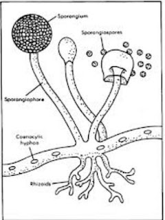
  • Less definition between cells; the hyphae of higher fungi have porous partitions called septa, which allow the passage of cytoplasm, organelles, and, sometimes, nuclei. Primitive fungi have few or no septa, so each organism is essentially a giant multinucleate supercell; these fungi are described as coenocytic.
  • Only the most primitive fungi, chytrids, have flagella.

eukaryotic cells

Other eukaryotic cells


Eukaryotes are a very diverse group, and their cell structures are equally diverse. Many have cell walls; many do not. Many have chloroplasts, derived from primary, secondary, or even tertiary endosymbiosis; and many do not. Some groups have unique structures, such as the cyanelles of the glaucophytes, the haptonema of the haptophytes, or the ejectisomes of the cryptomonads. Other structures, such as pseudopods, are found in various eukaryote groups in different forms, such as the lobose amoebozoans or the reticulose foraminiferans.

Uterus female reproductive system :blood supply

The uterus is supplied by arterial blood both from the uterine artery and the ovarian artery. Another anastomotic branch may also supply the uterus from anastomosis of these two arteries.

Nerve supply
Afferent nerves supplying uterus are T11 and T12. Sympathtic supply is from hypogastric plexus[disambiguation needed] and ovarian plexus. Parasympathetic supply is from second, third and fourth sacral nerves.

Development

The bilateral Müllerian ducts form during early fetal life. In males, MIF secreted from the testes leads to their regression. In females these ducts give rise to the Fallopian tubes and the uterus. In humans the lower segments of the two ducts fuse to form a single uterus, however, in cases of uterine malformations this development may be disturbed. The different uterine forms in various mammals are due to various degrees of fusion of the two Müllerian ducts.

Uterus female reproductive system :Position linea ligaments

The uterus is in the middle of the pelvic cavity in frontal plane (due to ligamentum latum uteri). The fundus does not surpass the linea terminalis, while the vaginal part of the cervix does not extend below interspinal line. The uterus is mobile and moves under the pressure of the full bladder or full rectum anteriorly, whereas if both are full it moves upwards. Increased intraabdominal pressure pushes it downwards. The mobility is conferred to it by musculo-fibrous apparatus that consists of suspensory and sustentacular part. Under normal circumstances the suspensory part keeps the uterus in anteflexion and anteversion (in 90% of women) and keeps it "floating" in the pelvis. The meaning of these terms are described below:
Distinction                         More common                   Less common
Position tipped           "Anteverted": Tipped forward     "Retroverted": Tipped backwards
Position of fundus     "Anteflexed": Fundus is pointing forward relative to the cervix     "Retroflexed": Fundus is pointing backwards


Sustentacular part supports the pelvic organs and comprises the larger pelvic diaphragm in the back and the smaller urogenital diaphragm in the front.

The pathological changes of the position of the uterus are:

    retroversion/retroflexion, if it is fixed
    hyperanteflexion - tipped too forward; most commonly congenital, but may be caused by tumors
    anteposition, retroposition, lateroposition - the whole uterus is moved; caused by parametritis or tumors elevation, descensus, prolaps  rotation (the whole uterus rotates around its longitudinal axis), torsion (only the body of the uterus rotates around) inversion

In cases where the uterus is "tipped", also known as retroverted uterus, women may have symptoms of pain during sexual intercourse, pelvic pain during menstruation, minor incontinence, urinary tract infections, difficulty conceiving, and difficulty using tampons. A pelvic examination by a doctor can determine if a uterus is tipped.

Uterus female reproductive system :Major ligaments

It is held in place by several peritoneal ligaments, of which the following are the most important (there are two of each):
Name                                    From                            To
Uterosacral ligament     Posterior cervix     Anterior face of sacrum
Cardinal ligaments     Side of the cervix     Ischial spines
Pubocervical ligament     Side of the cervix     Pubic symphysis

Uterus female reproductive system : Axes

Axes

Normally the uterus lies in anteversion & anteflexion. Anteversion is a forward angle between the axis of the cervix and that of the vagina measuring about 90 degrees, provided the urinary bladder and the rectum are empty. Anteflexion is a forward angle between the body and cervix at the isthmus measuring about 125 degrees, provided the bladder and rectum are empty. Uterus assumes anteverted position in 50% women, retorverted position in 25% women and rest have midposed uterus.

Uterus female reproductive system : Support

 Uterus female reproductive system : Support

The uterus is primarily supported by the pelvic diaphragm, perineal body and the urogenital diaphragm. Secondarily, it is supported by ligaments and the peritoneum (broad ligament of uterus)

Uterus female reproductive system Layers : Perimetrium

Perimetrium

    The loose connective tissue around the uterus.

Uterus female reproductive system Layers : Myometrium

Myometrium

    The uterus mostly consists of smooth muscle, known as "myometrium." The innermost layer of myometrium is known as the junctional zone, which becomes thickened in adenomyosis.
Perimetrium

Uterus female reproductive system Layers : Endometrium

Endometrium
    The lining of the uterine cavity is called the "endometrium". It consists of the functional endometrium and the basal endometrium from which the former arises. Damage to the basal endometrium results in adhesion formation and/or fibrosis (Asherman's syndrome). In all placental mammals, including humans, the endometrium builds a lining periodically which is shed or reabsorbed if no pregnancy occurs. Shedding of the functional endometrial lining is responsible for menstrual bleeding (known colloquially as a "period" in humans, with a cycle of approximately 28 days, +/-7 days of flow and +/-21 days of progression) throughout the fertile years of a female and for some time beyond. Depending on the species and attributes of physical and psychological health, weight, environmental factors of circadian rhythm, photoperiodism (the physiological reaction of organisms to the length of day or night), the effect of menstrual cycles to the reproductive function of the uterus is subject to hormone production, cell regeneration and other biological activities. The menstrual cycles may vary from a few days to six months, but can vary widely even in the same individual, often stopping for several cycles before resuming. Marsupials and monotremes do not have menstruation.

Uterus female reproductive system :Layers

There are three type of Layers


  1. Endometrium
  2. Myometrium
  3. Perimetrium

Uterus female reproductive system : Regions

From outside to inside, the path to the uterus is as follows:


    Cervix uteri - "neck of uterus"
  •         External orifice of the uterus
  •         Canal of the cervix
  •         Internal orifice of the uterus

    corpus uteri - "Body of uterus"
  •         Cavity of the body of the uterus
  •         Fundus (uterus)

Uterus Anatomy

Uterus Anatomy
'
The uterus is located inside the pelvis immediately dorsal (and usually somewhat rostral) to the urinary bladder and ventral to the rectum. The human uterus is pear-shaped and about 3 in. (7.6 cm) long, 4.5 cm broad (side to side) and 3.0 cm thick (anteroposterior). A nonpregnant adult uterus weighs about 60 grams. The uterus can be divided anatomically into four segments: The fundus, corpus, cervix and the internal os.

Formation Of Uterus In Mammals

Uterus Forms in mammals


Duplex  
There are two wholly separate uteri, with one fallopian tube each. Found in marsupials (such as kangaroos, Tasmanian devils, opossums, etc.), rodents (such as mice, rats, and guinea pigs), and lagomorpha (rabbits and hares).
Bipartite  
The two uteri are separate for most of their length, but share a single cervix. Found in ruminants (deer, moose, elk etc.), and cats.
Bicornuate 
The upper parts of the uterus remain separate, but the lower parts are fused into a single structure. Found in dogs, pigs, elephants, whales, dolphins, and prosimian primates among others.
Simplex  
The entire uterus is fused into a single organ. Found in higher primates (including humans and chimpanzees) . Occasionally, some individual females (including humans) may have a bicornuate uterus, a uterine malformation where the two parts of the uterus fail to fuse completely during fetal development.
In monotremes such as the platypus, the uterus is duplex and rather than nurturing the embryo, secretes the shell around the egg. It is essentially identical with the shell gland of birds and reptiles, with which the uterus is homologous.

Function Of Uterus

Function Of Uterus


The uterus consists of a body and a cervix. The cervix protrudes into the vagina. The uterus is held in position within the pelvis by condensations of endopelvic fascia, which are called ligaments. These ligaments include the pubocervical, transverse. cervical ligaments cardinal ligaments, and the uterosacral ligaments. It is covered by a sheet-like fold of peritoneum, the broad ligament.
The uterus is essential in sexual response by directing blood flow to the pelvis and to the external genitalia, including the ovaries, vagina, labia, and clitoris.
The reproductive function of the uterus is to accept a fertilized ovum which passes through the utero-tubal junction from the fallopian tube. It implants into the endometrium, and derives nourishment from blood vessels which develop exclusively for this purpose. The fertilized ovum becomes an embryo, attaches to a wall of the uterus, creates a placenta, and develops into a fetus (gestates) until childbirth. Due to anatomical barriers such as the pelvis, the uterus is pushed partially into the abdomen due to its expansion during pregnancy. Even during pregnancy the mass of a human uterus amounts to only about a kilogram (2.2 pounds).

Definition Of Uterus

Definition 
"Hystera" and "Uterine" redirect here. For the state of mind, see hysteria. For twins born of different fathers, see Uterine siblings.
The uterus (from Latin "uterus", plural uteri) or womb is a major female hormone-responsive reproductive sex organ of most mammals including humans. One end, the cervix, opens into the vagina, while the other is connected to one or both fallopian tubes, depending on the species. It is within the uterus that the fetus develops during gestation, usually developing completely in placental mammals such as humans and partially in marsupials such as kangaroos and opossums. Two uteri usually form initially in a female fetus, and in placental mammals they may partially or completely fuse into a single uterus depending on the species. In many species with two uteri, only one is functional. Humans and other higher primates such as chimpanzees, along with horses, usually have a single completely fused uterus, although in some individuals the uteri may not have completely fused. In English, the term uterus is used consistently within the medical and related professions, while the Germanic-derived term womb is more common in everyday usage.[citation needed]
Most animals that lay eggs, such as birds and reptiles, including most ovoviviparous species, have an oviduct instead of a uterus. Note however, that recent research into the biology of the viviparous (not merely ovoviviparous) skink Trachylepis ivensi has revealed development of a very close analogue to eutherian mammalian placental development.
In monotremes, mammals which lay eggs, namely the platypus and the echidnas, either the term uterus or oviduct is used to describe the same organ, but the egg does not develop a placenta within the mother and thus does not receive further nourishment after formation and fertilization.

Marsupials have two uteri, each of which connect to a lateral vagina and which both use a third, middle "vagina" which functions as the birth canal. Marsupial embryos form a choriovitelline "placenta" (which can be thought of as something between a monotreme egg and a "true" placenta), in which the egg's yolk sac supplies a large part of the embryo's nutrition but also attaches to the uterine wall and takes nutrients from the mother's bloodstream.

Uterus,female reproductive system

Uterus,female reproductive system


"Hystera" and "Uterine" redirect here. For the state of mind, see hysteria. For twins born of different fathers, see Uterine siblings.
The uterus (from Latin "uterus", plural uteri) or womb is a major female hormone-responsive reproductive sex organ of most mammals including humans. One end, the cervix, opens into the vagina, while the other is connected to one or both fallopian tubes, depending on the species. It is within the uterus that the fetus develops during gestation, usually developing completely in placental mammals such as humans and partially in marsupials such as kangaroos and opossums. Two uteri usually form initially in a female fetus, and in placental mammals they may partially or completely fuse into a single uterus depending on the species. In many species with two uteri, only one is functional. Humans and other higher primates such as chimpanzees, along with horses, usually have a single completely fused uterus, although in some individuals the uteri may not have completely fused. In English, the term uterus is used consistently within the medical and related professions, while the Germanic-derived term womb is more common in everyday usage.[citation needed]
Most animals that lay eggs, such as birds and reptiles, including most ovoviviparous species, have an oviduct instead of a uterus. Note however, that recent research into the biology of the viviparous (not merely ovoviviparous) skink Trachylepis ivensi has revealed development of a very close analogue to eutherian mammalian placental development.
In monotremes, mammals which lay eggs, namely the platypus and the echidnas, either the term uterus or oviduct is used to describe the same organ, but the egg does not develop a placenta within the mother and thus does not receive further nourishment after formation and fertilization.

Marsupials have two uteri, each of which connect to a lateral vagina and which both use a third, middle "vagina" which functions as the birth canal. Marsupial embryos form a choriovitelline "placenta" (which can be thought of as something between a monotreme egg and a "true" placenta), in which the egg's yolk sac supplies a large part of the embryo's nutrition but also attaches to the uterine wall and takes nutrients from the mother's bloodstream.